

Tiny Gold Particles can help early detection of Parkinson’s Disease
By- Jyoti Rawat
Tiny particles of the yellow metal, gold, may hold the key to a nanotechnology-based tool for the early detection of Parkinson’s Disease (PD). Parkinson’s disease is one of the fastest-growing neurological disorders globally. With an aging population and increasing life expectancy, the number of affected individuals in India is projected to rise significantly, placing immense pressure on healthcare systems. However, most diagnoses occur only after substantial neurodegeneration has already occurred.
Researchers are therefore seeking methods to detect the disease at an early stage, enabling appropriate measures to manage it effectively.
A group discussion among scientists at the Institute of Nano Science and Technology (INST), Mohali—an autonomous institute under the Department of Science and Technology (DST)—exploring how proteins behave differently in the brain during disease, sparked a promising idea. They began investigating whether they could assess a protein’s danger level by detecting its surface charge.
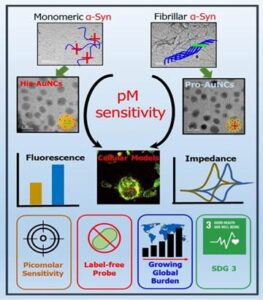
Their focus turned to a protein called α-synuclein, which is linked to PD. This protein undergoes a shape change, starting as a harmless form and eventually clumping into toxic aggregates that damage brain cells. The team set out to develop a sensor capable of distinguishing these protein forms based on their charge.
Their solution emerged in the form of gold nanoclusters (AuNCs), ultrasmall, glowing particles just a few nanometers in size. By coating these nanoclusters with naturally occurring amino acids, the researchers imparted selective “stickiness.” Proline-coated clusters were attracted to the normal protein form, while histidine-coated ones bound to the toxic aggregates. This approach successfully differentiated between the harmless monomeric form and the toxic aggregated (amyloid) form.
The journey from concept to proof-of-principle involved a wide range of experiments. The team started by engineering and purifying two forms of the α-synuclein protein (normal and mutant). They then synthesized amino acid–capped gold nanoclusters and characterized them using advanced techniques such as UV-Vis spectroscopy, fluorescence imaging, electron microscopy (TEM), and X-ray photoelectron spectroscopy (XPS) to understand their optical and structural properties. The interactions between nanoclusters and proteins were analyzed through gel electrophoresis, fluorescence quenching, and electrochemical methods like cyclic voltammetry and impedance spectroscopy, which enabled the team to measure the nanoclusters’ sensitivity in detecting different protein forms. Finally, the system was tested in human-derived SH-SY5Y neuroblastoma cells to confirm its safety and effectiveness in biological conditions.
The project was led by Dr. Sharmistha Sinha