
Microscopic Videos That Show the Invisible Wonders Around Us

The winners of this year’s Nikon Small World in Motion competition showcase the wiggly, feathery, undulating beauty of invisible life.

Inside us, below us, all around us, life abounds — but it’s often invisible to us. Only through the use of increasingly advanced microscopes can it be brought to our eyes in all its shimmery, glowing glory.
But capturing these minuscule moments requires the precision of a scientist and the patience of a photographer. “I see myself as a ranger in the African bush,” said Benedikt Pleyer, the third-place winner of a video contest that celebrates the dizzying beauty of the microscopic, the Nikon Small World in Motion competition. “You go out and see where the lions are, except they’re tiny and right in front of you.”
The Small World competition first began in 1975, when it accepted only photography. In 2011, a new and separate contest was created for moving images. This year’s judging panel included award-winning scientists, designers and directors of scientific institutions.
The winners, announced on Wednesday, reveal the most beautiful details that we can’t usually see — and how much we have to learn about the world around us.
First Place: Teeny Beating “Hairs”
Abnormal beating of airway cilia from a child with a disease called primary ciliary dyskinesia. Cilia are about 1/100th the width of a human hair.CreditCredit…Dr. Ning Xu
In airways like the lungs, throat and nostrils, cilia are tiny, hairlike structures that beat in coordinated waves to sweep away mucus. In a healthy person, cilia beat regularly and rapidly, but disease can change those rhythms. In his first-place video, Ning Xu, an optical engineer at the National University of Singapore, showed the cilia of a child with primary ciliary dyskinesia, a respiratory disorder that causes cilia to become weak, uncoordinated and unable to do their job. This failure can result in lifelong respiratory infections like bronchitis and pneumonia. “It’s not just cool optics,” Dr. Xu said. He is hoping that his work can help doctors better understand the disease, which affects an estimated one in 7,500 people worldwide.
Second Place: A Nosy Roundworm
A roundworm (nematode) and a single-celled Dileptus explore their microscopic world. A bundle of green and orange algae also hang out in view.CreditCredit…Nguyen Nam Nhat
Nguyen Nam Nhat was trying to film a single-celled freshwater predator called Dileptus when he noticed a contaminant in his sample: a pesky roundworm. “At first, I paid little attention to it,” said Mr. Nam Nhat, who studies the reproductive behaviors of microbes in Ho Chi Minh City, Vietnam. Not long after, though, he watched as the parasitic worm grabbed the Dileptus, which then had to sacrifice part of its taillike extremity to escape the roundworm’s clutches.
After examining similar cases, Mr. Nam Nhat found that, for roundworms, immobilizing competing organisms seems to be a typical defensive mechanism. It was entirely a chance observation made during filming, but for Mr. Nam Nhat, it highlighted the “unexpected interactions that can occur between microorganisms.”
Third Place: Smallest Jelly Aquarium
Jellyfish larvae, often called “sea lice” despite not being related to lice at all, are no larger than a grain of pepper. Here, a single larva is suspended in a water droplet.CreditCredit…Benedikt Pleyer
Bringing the microscopic alive for our honking human eyes is no easy feat. “When you shoot small creatures, people don’t have the feeling of scale in any conceivable way,” said Mr. Pleyer, a freelance microbe researcher based in Kyoto, Japan. Determined to find a workaround, he decided to use water droplets to better capture the dimensions of what he was seeing.
It took him years to figure out how: by pipetting microbes (in this case, jellyfish larvae, which are no larger than a grain of pepper) into a droplet suspended by a string. The Cassiopeia jellyfish shown here are armed with toxin-filled harpoons, fired when stressed. “It’s the smallest aquarium in the world,” Mr. Pleyer said.
Honorable Mentions: Dazzling Fungi, a Rainbow Sea Slug, Spiraling Algae and a Pulsing Heart Cell
A Penicillium species from which antibiotics emerge. Penicillium means “little brush” in Latin.CreditCredit…Wim van Egmond
When observed with the naked eye, most fungi “look like unpleasant gray or brown fluff,” said Wim van Egmond, who began making time-lapse videos more than 12 years ago while working at the Micropia Museum in Amsterdam. Maybe you know fungi best as mushrooms, or that they help humans by producing antibiotics. In this time-lapse, you can see a Penicillium species forming a thick carpetlike network called a mycelium, from which emerge bright red droplets full of antibiotics and other substances. “When you get to know them better,” Mr. van Egmond said of the fungi in these close-up views, “you will hopefully get more appreciation for these modest beings.”
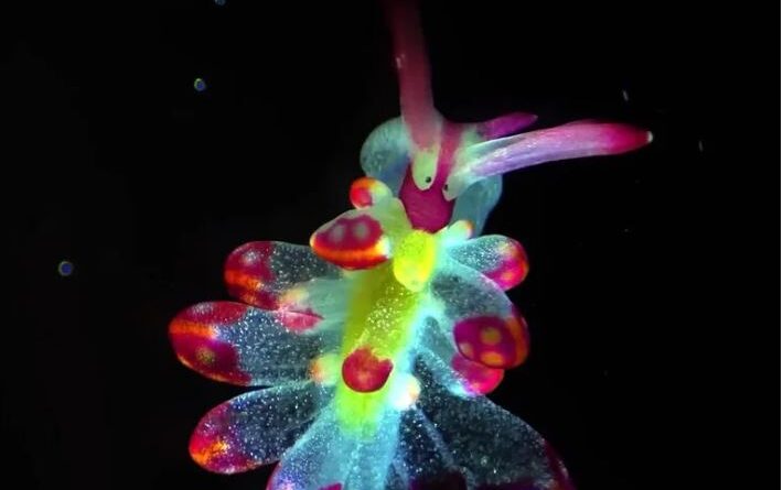
Cyerce kikutarobabai, better known as the butterfly sea slug, is a colorful creature covered in winglike structures that help it breathe and swim — and give the appearance of butterfly wings. Igor Adameyko of the Center for Brain Research at the Medical University of Vienna said that observing the 4-millimeter-long sea slug allowed him to appreciate that the structures were not simply decorative. “It was one of those moments,” he said, “when scientific observation and visual beauty became inseparable.”
These freshwater algae, Volvox aureus, are found all over the place, most commonly in ponds, lakes and rivers. In the sample collected and captured by Gerd Günther, a photomicrographer based in Düsseldorf, Germany, the colonies contain something called zygospores, which represent the algae’s sexual resting stage. Zygospores act as a biological time capsule to preserve the algae’s genetic code when conditions deteriorate, such as amid a steep drop in temperature.